
Renal Cell Carcinoma
Staging
As with most cancers, identifying the stage of RCC is essential for determining the prognosis (likely outcome) and for planning treatment. Staging helps doctors describe where the cancer is located, whether it has spread and whether other organs in the body are affected by it.
RCC may be staged in two ways. First, your doctor will evaluate the results of your physical exam and imaging tests and determine a clinical stage. Then, if surgery is performed, a pathologist will examine tissue taken from the tumor and nearby lymph nodes and assign a pathologic stage, which provides much more detail about the cancer. The enhanced detail of the pathologic stage is important to determine the best treatment options and to predict the prognosis; however, sometimes the clinical staging provides sufficient information to guide therapy.
RCC is classified according to the tumor, node and metastasis (TNM) system developed by the American Joint Committee on Cancer (AJCC) (Table 1). Doctors categorize the tumor (T) according to its size and location, whether cancer cells are found in nearby lymph nodes (N) and whether the cancer has metastasized (M) – or spread – to other parts of the body. Once the RCC has been classified with the TNM system, an overall stage is assigned (Table 2). RCC is staged from Stage I to Stage IV. If the RCC is diagnosed as Stage I, II or III, it is non-metastatic or localized, meaning it hasn’t metastasized, or spread, to other parts of the body. Stage IV RCC is considered metastatic and has spread beyond the kidney and surrounding lymph nodes.
The Fuhrman grade of cancer is also an important tool in assessing RCC. This grade refers to how closely the cancer cells resemble normal cells under the microscope. The grading scale ranges from 1 to 4, with 1 representing cancer cells that look similar to normal cells and 4 representing cancer cells that look vastly different from normal cells. The lower the grade, the better the outlook is for the person with RCC.
Table 1. AJCC TNM Classification for Kidney Cancer
| Classification | Definition |
| Tumor (T) | |
| TX | Primary tumor cannot be assessed. |
| T0 | No evidence of primary tumor. |
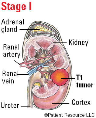
| T1 | Tumor ≤ (less than or equal to) 7 cm in greatest dimension, limited to the kidney. |
| T1a | Tumor ≤ (less than or equal to) 4 cm in greatest dimension, limited to the kidney. |
| T1b | Tumor > (more than) 4 cm but ≤ (less than or equal to) 7 cm in greatest dimension, limited to the kidney. |
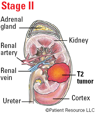
| T2 | Tumor > (more than) 7 cm in greatest dimension, limited to the kidney. |
| T2a | Tumor > (more than) 7 cm but ≤ (less than or equal to) 10 cm in greatest dimension, limited to the kidney. |
| T2b | Tumor > (more than) 10 cm, limited to the kidney. |
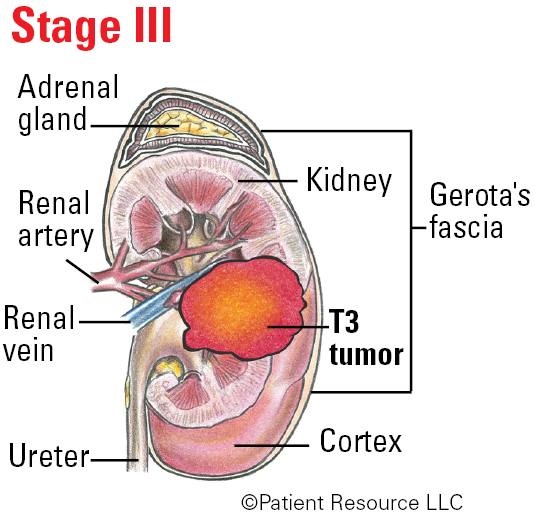
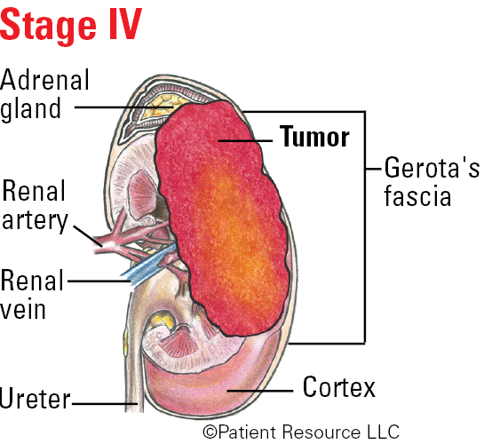
| T3 | Tumor extends into major veins or perinephric tissues, but not into the ipsilateral adrenal gland (a small gland on top of each kidney) and not beyond Gerota’s fascia (an envelope of tissue that surrounds the kidney). |
| T3a | Tumor extends into the renal vein (the large blood vessel leaving the kidney) or its segmental branches, or invades the pelvicalyceal system, or invades perirenal (surrounding the kidney) and/or renal sinus (within the kidney) fat but not beyond Gerota’s fascia. |
| T3b | Tumor extends into the vena cava (the large vein leaving the heart) below the diaphragm (the muscle under the lungs that helps with breathing). |
| T3c | Tumor extends into the vena cava above the diaphragm or invades the wall of the vena cava. |
| T4 | Tumor invades beyond Gerota’s fascia (including contiguous extension into the ipsilateral adrenal gland). |
| Node (N) | |
| NX | Regional lymph nodes cannot be assessed. |
| N0 | No regional lymph node metastasis. |
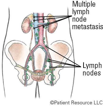
| N1 | Metastasis in regional lymph node(s). |
| Metastasis (M) | |
| M0 | No distant metastasis. |
| M1 | Distant metastasis. |
Table 2: Stages of RCC
| Stage | TNM Classifications |
| Stage I | T1, N0, M0 |
| Stage II | T2, N0, M0 |
| Stage III |
T1 or T2, N1, M0
T3, N0 or N1, M0 |
| Stage IV |
T4, Any N, M0
Any T, Any N, M1 |
Illustrated Stages of Kidney Cancer
Metastatic RCC
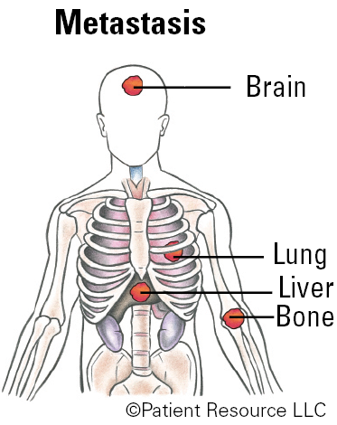
If the RCC is diagnosed as Stage IV, it is metastatic, which means it has spread from the kidney to another part of the body (called a “metastatic site”). The most common metastatic sites for RCC are the lungs, lymph nodes, bones, brain and liver.
Metastatic tumors have the same type of abnormal cells as the primary (original) tumor and are referred to by the same name. So, if RCC metastasizes to the liver, for example, the cancer cells in the liver are still RCC cells and the disease is called metastatic RCC rather than liver cancer. The metastatic disease is also treated as RCC, not as liver cancer.
Throughout the diagnosis and staging processes, talk openly with your health care team. Ask any questions you may have so you can better understand your specific cancer and the best options for you.
