
Uterine (Endometrial) Cancer
Overview and Staging
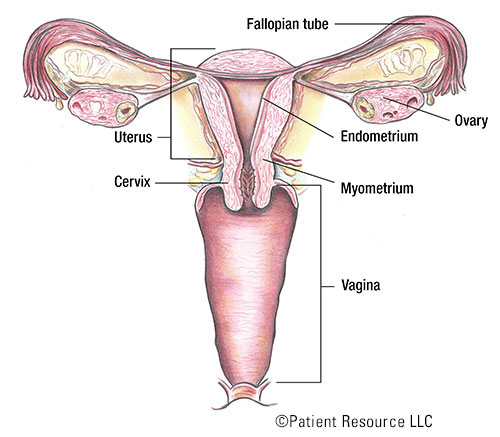
The uterus is a hollow organ about the size and shape of a pear. When a woman is pregnant, the fetus grows in the uterus, which is also referred to as the womb. The uterus has two main parts: the cervix, which is the “neck” of the uterus that extends into the vagina, and the upper part, or the body or corpus of the uterus.
The main type of cancer of the uterus is called endometrial cancer. It starts in the cells of the endometrium, the inner lining of the uterus. Endometrial cancer is the most common gynecologic cancer. Although the cervix is part of the uterus, cancer that occurs in the cervix is called cervical cancer and is not the same as endometrial cancer.
Diagnosing and staging
The results of an endometrial biopsy allow your doctor to diagnose endometrial cancer. After cancer has been diagnosed, it is staged. In staging cancer, your doctor determines the extent of the cancer based on the size of the tumor and whether the cancer has spread and, if so, how far.
Endometrial cancer is often staged based on the surgical removal of the tumor. To establish whether the cancer cells have spread, the surgeon may also remove nearby lymph nodes, tissues and fluid within the pelvic and/or abdominal cavity. A pathologist’s examination of these samples and information from imaging tests help the doctor determine an accurate and exact stage, which is the most important factor in choosing an appropriate treatment option.
Like many other cancer types, endometrial cancer is classified according to the tumor, node, metastasis (TNM) system developed by the American Joint Committee on Cancer (AJCC). The tumor (T) is analyzed and categorized based on its size and location, whether cancer cells occupy nearby lymph nodes (N), and whether cancer cells have metastasized (M) or spread to other organs and body parts. Once an endometrial cancer has been classified with this system, an overall stage is assigned (see Staging Illustrations).
Table 1. TNM Uterine Cancer Classifications
| Classification | Definition |
| Tumor (T) | |
| TX | Primary tumor cannot be assessed. |
| T0 | No evidence of primary tumor. |
| T1 | Tumor confined to the corpus uteri (body of the uterus), including endocervical glandular involvement. |
| T1a | Tumor limited to the endometrium (inner lining of the uterus) or invading less than half the myometrium (muscular layer of the uterus). |
| T1b | Tumor invading one half or more of the myometrium (muscular layer of the uterus). |
| T2 | Tumor invading the stromal connective tissue of the cervix but not extending beyond the uterus. Does not include endocervical glandular involvement. |
| T3 | Tumor involving serosa (outer smooth layer of the uterus), adnexa (area connecting to the uterus such as fallopian tubes and ovaries), vagina or parametrium (layer of tissue that separates the cervix from the bladder). |
| T3a | Tumor involving the serosa (outer smooth layer of the uterus) and/or adnexa (area connecting to the uterus such as fallopian tubes and ovaries) (direct extension or metastasis). |
| T3b | Vaginal involvement (direct extension or metastasis) or parametrial (layer of tissue that separates the cervix from the bladder) involvement. |
| T4 | Tumor invading the bladder mucosa (lining) and/or bowel. |
| Node (N) | |
| NX | Regional lymph nodes cannot be assessed. |
| N0 | No regional lymph node metastasis. |
| N0(i+) | Isolated tumor cells in regional lymph node(s) no greater than 0.2 mm. |
| N1 | Regional lymph node metastasis to pelvic lymph nodes. |
| N1mi | Regional lymph node metastasis (greater than 0.2 mm but not greater than 2.0 mm in diameter) to pelvic lymph nodes. |
| N1a | Regional lymph node metastasis (greater than 2.0 mm in diameter) to pelvic lymph nodes. |
| N2 | Regional lymph node metastasis to para-aortic lymph nodes (lymph nodes along the aorta), with or without positive pelvic lymph nodes. |
| N2mi | Regional lymph node metastasis (greater than 0.2 mm but not greater than 2.0 mm in diameter) to para-aortic lymph nodes (lymph nodes along the aorta), with or without positive pelvic lymph nodes. |
| N2a | Regional lymph node metastasis (greater than 2.0 mm in diameter) to para-aortic lymph nodes (lymph nodes along the aorta), with or without positive pelvic lymph nodes. |
| Metastasis (M) | |
| M0 | No distant metastasis. |
| M1 | Distant metastasis (includes metastasis to inguinal lymph nodes, intraperitoneal disease, lung, liver or bone). (It excludes metastasis to pelvic or para-aortic lymph nodes (lymph nodes along the aorta), vagina, uterine serosa (outer smooth layer of the uterus), or adnexa (area connecting to the uterus such as fallopian tubes and ovaries). |
Table 2. Stages of Uterine Cancer
| Stage | TNM Classification |
| I | T1, N0, M0 |
| IA | T1a, N0, M0 |
| IB | T1b, N0, M0 |
| II | T2, N0, M0 |
| III | T3, N0, M0 |
| IIIA | T3a, N0, M0 |
| IIIB | T3b, N0, M0 |
| IIIC1 | T1 to T3, N1/N1mi/N1a, M0 |
| IIIC2 | T1 to T3, N2/N2mi/N2a, M0 |
| IVA | T4, Any N, M0 |
| IVB | Any T, Any N, M1 |
